

REVIEW ARTICLE
The role of computational pathology in predicting expression of selected immunohistochemical markers
1
IDEAS Research Institute, Warsaw, Poland
2
Maria Sklodowska-Curie National Research Institute of Oncology, Tumor Pathology Department, Gliwice, Poland
3
Faculty of Electrical Engineering, Warsaw University of Technology, Warsaw, Poland
Submission date: 2026-03-31
Acceptance date: 2026-05-02
Publication date: 2026-06-25
Corresponding author
Pol J Pathol 2026;77(2):209-215

KEYWORDS
TOPICS
ABSTRACT
Immunohistochemistry (IHC) remains a cornerstone of precision oncology, providing essential diagnostic, prognostic, and predictive molecular data. However, the traditional manual assessment of IHC slides faces persistent challenges regarding interobserver variability, reproducibility, and high diagnostic workloads. To address these limitations, artificial intelligence (AI) is increasingly integrated into computational pathology workflows. This paper outlines the current landscape of AI-assisted IHC, highlighting significant trends such as automated scoring, explainable AI, virtual staining, and multiplex analysis. We explore the transition from basic image analysis to advanced deep learning architectures capable of predicting specific IHC biomarker expression directly from standard hematoxylin and eosin morphology. Furthermore, we examine the commercial ecosystem of these tools and highlight the critical pre-analytical bottlenecks that hinder widespread clinical adoption.
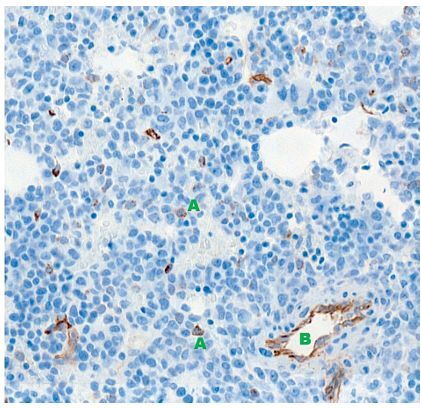
Finally, we present a practical case study demonstrating an automated, deep learning-
based pipeline for quantifying CD34-positive myeloblasts in acute myeloid leukemia and myelodysplastic syndromes. Ultimately, while AI holds vast potential to optimize turnaround times and streamline laboratory triage, its successful clinical implementation will depend on seamless integration into existing digital pathology platforms and standardization of pre-analytical variables, moving the field from fragmented algorithms to reliable, standard-of-care diagnostic tools.
REFERENCES (55)
1.
Coons AH, Creech HJ, Jones RN. Immunological properties of an antibody containing a fluorescent group. Proc Soc Exp Biol Med 1941; 47: 200-202.
2.
Ortiz Hidalgo C. Immunohistochemistry in historical perspective: knowing the past to understand the present. Methods Mol Biol 2022; 2422: 17-31.
3.
Nakane PK, Pierce GB Jr. Enzyme-labeled antibodies: preparation and application for the localization of antigens. J Histochem Cytochem 1966; 14: 929-931.
4.
Duraiyan J, Govindarajan R, Kaliyappan K, Palanisamy M. Applications of immunohistochemistry. J Pharm Bioallied Sci 2012; 4: S307-309.
5.
Chang TG, Park S, Schäffer AA, Jiang P, Ruppin E. Hallmarks of artificial intelligence contributions to precision oncology. Nat Cancer 2025; 6: 417-431.
6.
Ricci C, Ambrosi F, Franceschini T, Giunchi F, Maracci ME, Sirolli M, et al. H&E and OCT4/CD34 for the assessment of lympho-vascular invasion in seminoma and embryonal carcinoma. Pathol Res Pract 2023; 242: 154337.
7.
Flach R, Fransen N, Sonnen A, Nguyen T, Breimer G, Veta M, et al. Implementation of artificial intelligence in diagnostic practice as a next step after going digital: the UMC Utrecht perspective. Diagnostics (Basel) 2022; 12: 1042.
8.
Song AH, Jaume G, Williamson DFK, Lu MY, Vaidya A, Miller TR, et al. Artificial intelligence for digital and computational pathology. Nat Rev Bioeng 2023; 1: 930-949.
9.
Gu Q, Patel A, Hanna MG, Lennerz JK, Garcia C, Zarella M, et al. Bridging the clinical-computational transparency gap in digital pathology. Arch Pathol Lab Med 2025; 149: 276-287.
10.
Verghese G, Lennerz JK, Ruta D, Ng W, Thavaraj S, Siziopikou KP, et al. Computational pathology in cancer diagnosis, prognosis, and prediction – present day and prospects. J Pathol 2023; 260: 551-563.
11.
Fauzi MF, Pennell M, Sahiner B, Chen W, Shana’ah A, Hemminger J, et al. Classification of follicular lymphoma: the effect of computer aid on pathologists grading. BMC Med Inform Decis Mak 2015; 15: 115.
12.
Feng M, Deng Y, Yang L, Jing Q, Zhang Z, Xu L, et al. Automated quantitative analysis of Ki-67 staining and HE images recognition and registration based on whole tissue sections in breast carcinoma. Diagn Pathol 2020; 15: 65.
13.
Wang W, Gong Y, Chen B, Guo H, Wang Q, Li J, et al. Quantitative immunohistochemistry analysis of breast Ki67 based on artificial intelligence. Open Life Sci 2024; 19: 20221013.
14.
Dawe M, Shi W, Liu TY, Lajkosz K, Shibahara Y, Gopal NEK, et al. Reliability and variability of Ki-67 digital image analysis methods for clinical diagnostics in breast cancer. Lab Invest 2024; 104: 100341.
15.
Dy A, Nguyen NJ, Meyer J, Dawe M, Shi W, Androutsos D, et al. AI improves accuracy, agreement and efficiency of pathologists for Ki67 assessments in breast cancer. Sci Rep 2024; 14: 1283.
16.
Khameneh FD, Razavi S, Kamasak M. Automated segmentation of cell membranes to evaluate HER2 status in whole slide images using a modified deep learning network. Comput Biol Med 2019; 110: 164-174.
17.
Jung M, Song SG, Cho SI, Shin S, Lee T, Jung W, et al. Augmented interpretation of HER2, ER, and PR in breast cancer by artificial intelligence analyzer: enhancing interobserver agreement through a reader study of 201 cases. Breast Cancer Res 2024; 26: 31.
18.
Zannoni GF, Angelico G, d’Amati A, D’Alessandris N, Scaglione G, Urtueta BP, et al. Interobserver agreement in immunohistochemical evaluation of folate receptor alpha (FRα) in ovarian cancer: a multicentre study. Int J Mol Sci 2025; 26: 7687.
19.
Ghasemi A, Hashtarkhani S, Schwartz DL, Shaban-Nejad A. Explainable artificial intelligence in breast cancer detection and risk prediction: a systematic scoping review. Cancer Innov 2024; 3: e136.
20.
Gunning D, Aha D. DARPA’s explainable artificial intelligence (XAI) program. AI Mag 2019; 40: 44-58.
21.
Renner JA, Riley PC. Using machine learning for chemical-free histological tissue staining. J Histotechnol 2024; 47: 180-183.
22.
Ji C, Oshima K, Urata T, Kimura F, Ishii K, Uehara T, et al. Transformation from hematoxylin-and-eosin staining to Ki-67 immunohistochemistry digital staining images using deep learning: experimental validation on the labeling index. J Med Imaging (Bellingham) 2024; 11: 047501.
23.
Zhang Y, Işıl Ç, Yang X, Li Y, Elia A, Atlan K, et al. Deep learning-enabled virtual multiplexed immunostaining of labelfree tissue for vascular invasion assessment. BME Front 2026; 7: 0226.
24.
Klöckner P, Teixeira J, Montezuma D, Fraga J, Horlings HM, Cardoso JS, et al. H&E to IHC virtual staining methods in breast cancer: an overview and benchmarking. NPJ Digit Med 2025; 8: 384.
25.
Ghahremani P, Marino J, Hernandez-Prera J, de la Iglesia JV, Slebos RJ, Chung CH, et al. An AI-ready multiplex staining dataset for reproducible and accurate characterization of tumor immune microenvironment. Med Image Comput Comput Assist Interv 2023; 14225: 704-713.
26.
Lim JCT, Yeong JPS, Lim CJ, Ong CCH, Wong SC, Chew VSP, et al. An automated staining protocol for seven-colour immunofluorescence of human tissue sections for diagnostic and prognostic use. Pathology 2018; 50: 333-341.
27.
Ghahremani P, Li Y, Kaufman A, Vanguri R, Greenwald N, Angelo M, et al. Deep learning-inferred multiplex immunofluorescence for immunohistochemical image quantification. Nat Mach Intell 2022; 4: 401-412.
28.
Tan WCC, Nerurkar SN, Cai HY, Ng HHM, Wu D, Wee YTF, et al. Overview of multiplex immunohistochemistry/immunofluorescence techniques in the era of cancer immunotherapy. Cancer Commun (Lond) 2020; 40: 135-153.
29.
Akbarnejad A, Ray N, Barnes P, Bigras G. Toward accurate deep learning-based prediction of Ki67, ER, PR, and HER2 status from H&E-stained breast cancer images. Appl Immunohistochem Mol Morphol 2025; 33: 131-141.
30.
Gamble P, Jaroensri R, Wang H, Tan F, Moran M, Brown T, et al. Determining breast cancer biomarker status and associated morphological features using deep learning. Commun Med (Lond) 2021; 1: 14.
31.
Liu Y, Li X, Zheng A, Zhu X, Liu S, Hu M, et al. Predict Ki-67 positive cells in H&E-stained images using deep learning independently from IHC-stained images. Front Mol Biosci 2020; 7: 183.
32.
Jiao P, Zheng Q, Yang R, Ni X, Wu J, Chen Z, et al. Prediction of HER2 status based on deep learning in H&E-stained histopathology images of bladder cancer. Biomedicines 2024;.
33.
L1i2u: S1,5 Z83h.a ng B, Liu Y, Han A, Shi H, Guan T, et al. Unpaired stain transfer using pathology-consistent constrained generative adversarial networks. IEEE Trans Med Imaging 2021; 40:.
34.
1B9is7w7a-s1 9T8, 9S.u zuki H, Ishikawa M, Kobayashi N, Obi T. Generative adversarial network based digital stain conversion for generating RGB EVG stained image from hyperspectral H&E stained image. J Biomed Opt 2023; 28: 056501.
35.
Yan R, He Q, Liu Y, Ye P, Zhu L, Shi S, et al. Unpaired virtual histological staining using prior-guided generative adversarial networks. Comput Med Imaging Graph 2023; 105: 102185.
36.
Lysik M, Swiderska Z, Markiewicz T, Cierniak S, Lorent M. HE-to-PAS histological stain conversion by GAN in renal pathology. In: 2022 International Joint Conference on Neural Networks (IJCNN). IEEE; 2022: 1-7.
37.
Das A, Tomita N, Syme KJ, Ma W, O’Connor P, Corbett KN, et al. Cross-modality learning for predicting immunohistochemistry biomarkers from hematoxylin and eosin-stained whole slide images. Am J Pathol 2025; 195: 2400-2410.
38.
Pantanowitz L, Sharma A, Carter AB, Kurc T, Sussman A, Saltz J. Twenty years of digital pathology: an overview of the road travelled, what is on the horizon, and the emergence of vendorneutral archives. J Pathol Inform 2018; 9: 40.
39.
Limaiem F. Artifacts in routine histopathology: unveiling potential causes of misdiagnosis. J Clin Med Images Case Rep 2024; 4: 1.
40.
Mebratie DY, Dagnaw GG. Review of immunohistochemistry techniques: applications, current status, and future perspectives. Semin Diagn Pathol 2024; 41: 154-160.
41.
Scalia CR, Boi G, Bolognesi MM, Riva L, Manzoni M, DeSmedt L, et al. Antigen masking during fixation and embedding, dissected. J Histochem Cytochem 2017; 65: 5-20.
42.
Gestewitz M, Praetorius M, Albrecht T. Decalcification method for nondestructive exposure of human inner ear specimens using using ultrasonic EDTA scrubbing. Biotech Histochem 2022; 97: 347-354.
43.
Orazi A. Histopathology in the diagnosis and classification of acute myeloid leukemia, myelodysplastic syndromes, and myelodysplastic/myeloproliferative diseases. Pathobiology 2007; 74: 97-114.
44.
Elmore JG, Longton GM, Carney PA, Geller BM, Onega T, Tosteson AN, et al. Diagnostic concordance among pathologists interpreting breast biopsy specimens. JAMA 2015; 313: 1122-1132.
45.
Van der Wel MJ, Coleman HG, Bergman JJGHM, Jansen M, Meijer SL, et al. Histopathologist features predictive of diagnostic concordance at expert level among a large international sample of pathologists diagnosing Barrett’s dysplasia using digital pathology. Gut 2020; 69: 811-822.
46.
Rakha EA, Quinn CM, Provenzano E, Pinder SE, Ellis IO. Diagnostic discordance and error in breast pathology: causes, classifications, and medicolegal implications. Mod Pathol 2026; 39: 100942.
47.
Abdelateef SM, Al-Rubaie HA, Abid SA. Immunohistochemical analysis of CD34 to evaluate angiogenesis in chronic lymphocytic leukemia. J Fac Med Baghdad 2013; 55: 131-134.
48.
Maschio LB, Madallozo BB, Capellasso BA, Jardim BV, Moschetta MG, Jampietro J, et al. Immunohistochemical investigation of the angiogenic proteins VEGF, HIF-1α and CD34 in invasive ductal carcinoma of the breast. Acta Histochem 2014; 116: 148-157.
49.
Jocher G, Qiu J. Ultralytics YOLO11. Ultralytics; 2024. Available from: https://github.com/ultralytics... (accessed: 31.03.2026).
50.
Carion N, Gustafson L, Hu YT, Debnath S, Hu R, Suris D, et al. SAM 3: segment anything with concepts. arXiv [Preprint] 2025.
51.
Tian Y, Ye Q, Doermann D. YOLO12: attention-centric realtime object detectors. arXiv preprint arXiv:2502.12524; 2025.
52.
Tian Y, Ye Q, Doermann D. YOLO12: attention-centric real-time object detectors. GitHub repository; 2025. Available from: https://github.com/sunsmarterj... (accessed: 31.03.2026).
53.
Greenberg PL, Cox C, LeBeau M, Fenaux P, Morel P, Sanz G, et al. International scoring system for evaluating prognosis in myelodysplastic syndromes. Blood 1997; 89: 2079-2088.
54.
Greenberg PL, Tuechler H, Schanz J, Sanz G, Garcia-Manero G, Solé F, et al. Revised international prognostic scoring system for myelodysplastic syndromes. Blood 2012; 120: 2454-2465.
55.
Landis JR, Koch GG. The measurement of observer agreement for categorical data. Biometrics 1977; 33: 159-174.
| eISSN: | 2084-9869 |
| ISSN: | 1233-9687 |

We process personal data collected when visiting the website. The function of obtaining information about users and their behavior is carried out by voluntarily entered information in forms and saving cookies in end devices. Data, including cookies, are used to provide services, improve the user experience and to analyze the traffic in accordance with the Privacy policy. Data are also collected and processed by Google Analytics tool (more).
You can change cookies settings in your browser. Restricted use of cookies in the browser configuration may affect some functionalities of the website.
You can change cookies settings in your browser. Restricted use of cookies in the browser configuration may affect some functionalities of the website.
